Quantifoil and the revolution of cryo-electron microscopy: a precision support for structural research
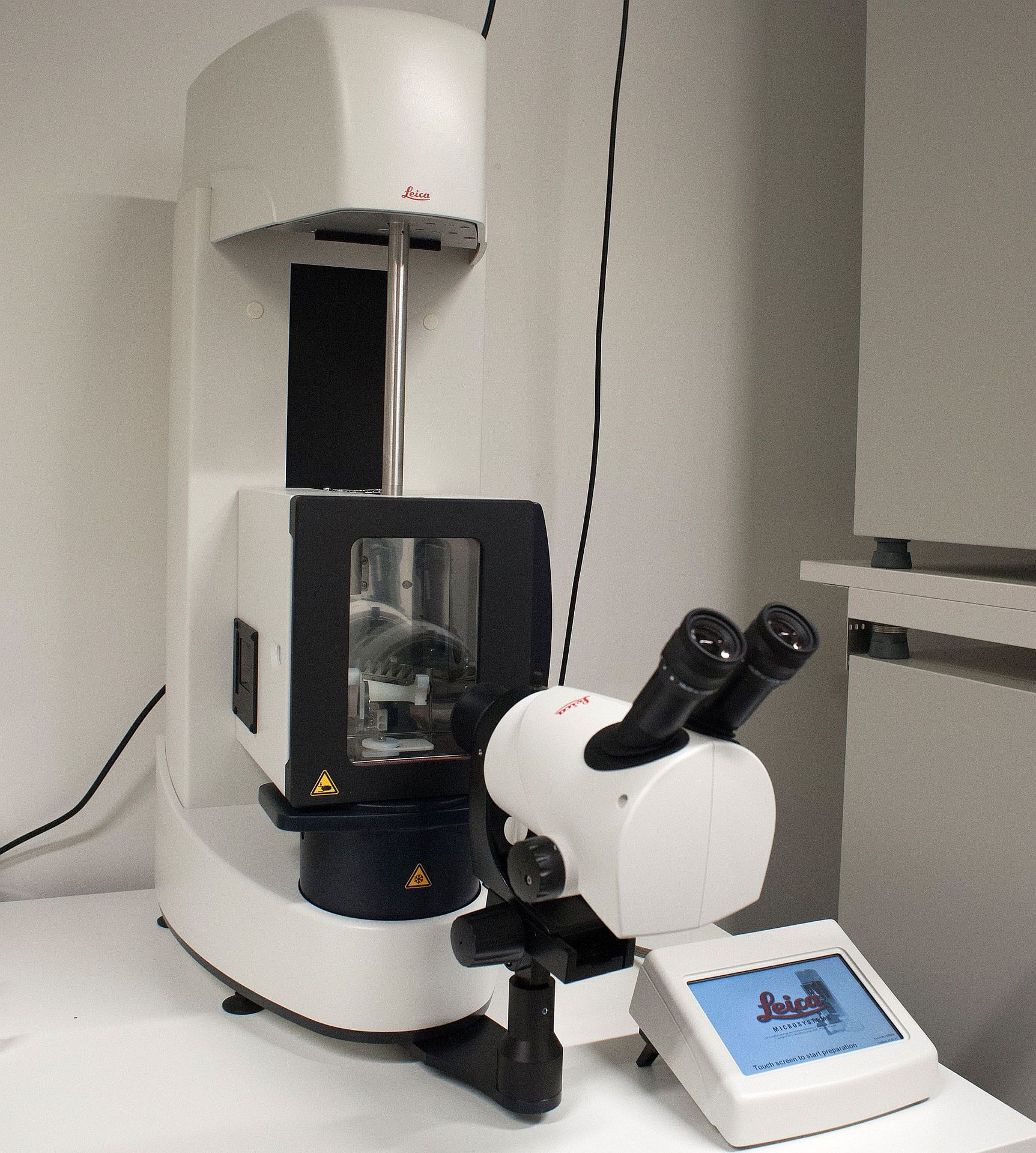
In the field of structural biology and nanoscience, cryo-electron microscopy (cryo-EM) has revolutionised the analysis of macromolecular structures. One of the key elements in this cutting-edge technique is the support used to immobilise the samples: the microscopy grid. Among all the options available on the market, Quantifoil® grids have established themselves as the global standard.
Why are Quantifoil grids so important?
Compatibility with the latest generation of microscopes
The Quantifoil grids are designed to be perfectly compatible with the most advanced electron microscopes.
Background noise reduction
The ultrathin amorphous carbon film used in Quantifoil grids reduces background noise and interference, which improves the signal-to-noise ratio, a crucial element for achieving atomic resolution.
Uniform vitrification
Thanks to the well-calibrated holes, the ice layer containing the sample is vitrified uniformly, without crystallisation, a crucial criterion for successful cryo-EM..
Applications of Quantifoil grids
The Quantifoil grids are used in many disciplines:
- Study of membrane proteins, ribosomal complexes or viruses
- Analysis of nanoparticles, polymers or exosomes
- Electron tomography and 3D reconstructions of biological complexes
- Correlative microscopy (CLEM) combining fluorescence and EM
The scientific contribution of cryo-EM
A recent publication published in Medicine/Sciences (avril 2021) available as a PDF here .
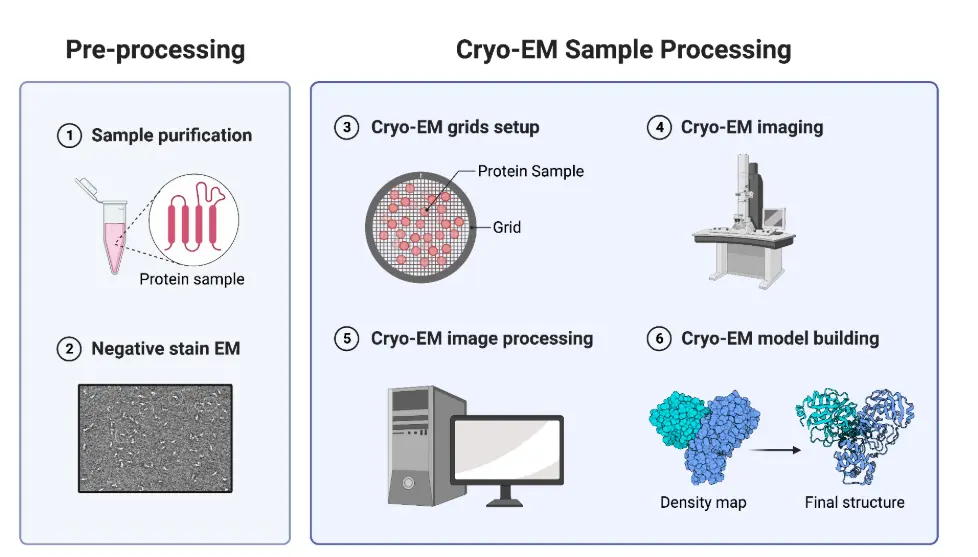
Typical Cryo-EM Workflow
- Purified protein shipped to the Cryo-Electron Microscopy Center (CEMC)
- Plunge freezing of the protein sample on a TEM grid
- Cryo grid screening and validation
- Data collection
- Remote data delivery
- Data analysis and 3D structure determination
Cryo-EM Methodology: Current Aspects and Future Directions
📚 By Radostin Danev, Haruaki Yanagisawa & Masahide Kikkawa – University of Tokyo
Cryo-electron microscopy (cryo-EM) continues to revolutionize structural biology. In a recent landmark article, researchers from the University of Tokyo explore the current methodologies and future directions of cryo-EM — a technology that’s reshaping how we understand biomolecular structures.
👉 Download the full article in PDF here:
Cryo-EM Methodology – Danev et al. (2021)
Mallax Pharma :
A Trusted Name in Modern Pharmaceutical Innovation
Mallax has become a leading force in pharmaceutical development, trusted for its commitment to innovation, patient care, and strategic partnerships. At the core of Mallax Pharma is a vision to make advanced medical solutions accessible and effective across the globe.
Whether you're searching for the latest in malax medicine, researching Sana Pharma collaborations, or exploring new biotech initiatives like Mpolux, Mallax stands out for quality and performance.
Why Mallax Is Gaining Global Recognition
The name Mallax has quickly grown to represent more than just a brand—it signifies trust, research excellence, and therapeutic reliability. Pharmaceutical professionals and healthcare providers consistently turn to Mallax Pharma for :
- High-efficacy formulations
- Regulatory-compliant product lines
- International partnerships and distribution
- Advanced R&D pipelines
As the demand for specialized pharmaceuticals grows, malax medicine offers solutions tailored to both hospital and outpatient needs.
Strategic
Our latest content
Check out what's new in our company !
Our latest content
Check out what's new in our company !